Diseases of the Retina

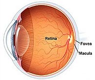
Retinal diseases specifically affect the retina — a layer of tissue at the back of the eye that is responsible for vision. These diseases can affect the retina, the macula (area of central vision), or the fovea at the center of the macula. Many retinal diseases share common symptoms and treatments, but each has unique characteristics.

Age-Related Macular Degeneration

Age-related macular degeneration (AMD) is one of the most common causes of poor vision after age 60. AMD is a deterioration or breakdown of the macula. The macula is a small area at the center of the retina in the back of the eye that allows us to see fine details clearly and perform activities such as reading and driving.

The visual symptoms of AMD involve loss of central vision. While peripheral (side) vision is unaffected, with AMD, one loses the sharp, straight-ahead vision necessary for driving, reading, recognizing faces, and looking at detail.

Although the specific cause is unknown, AMD seems to be part of aging. While age is the most significant risk factor for developing AMD, heredity, blue eyes, high blood pressure, cardiovascular disease, and smoking have also been identified as risk factors. AMD accounts for 90% of new cases of legal blindness in the United States.

Nine out of 10 people who have AMD have atrophic or “ dry” AMD, which results in thinning of the macula. Dry AMD takes many years to develop. A specific vitamin regimen has been shown to slow progression of dry AMD.

Exudative or “ wet” AMD is less common (occurring in one out of 10 people with AMD) but is more serious. In the wet form of AMD, abnormal blood vessels may grow in a layer beneath the retina, leaking fluid and blood and creating distortion or a large blind spot in the center of your vision. If the blood vessels are not growing directly beneath the macula, laser surgery is usually the treatment of choice. The procedure usually does not improve vision but tries to prevent further loss of vision. For those patients with wet AMD whose blood vessels are growing directly under the center of the macula, a procedure called photodynamic therapy (PDT), which causes fewer visual side effects, is sometimes used. Intravitreal injections of certain medications can also be used in these cases.

Back to Top

Nonproliferative Diabetic Retinopathy

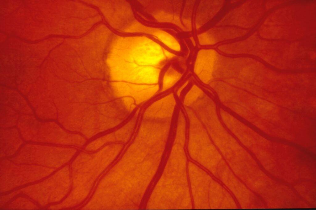
If you have diabetes mellitus, your body does not use and store glucose properly. Over time, diabetes can damage blood vessels in the retina, the nerve layer at the back of the eye that senses light and helps to send images to the brain. The damage to retinal vessels is referred to as diabetic retinopathy.

Nonproliferative diabetic retinopathy (NPDR), commonly known as background retinopathy, is an early stage of diabetic retinopathy. In this stage, tiny blood vessels within the retina leak blood or fluid. The leaking fluid causes the retina to swell or to form deposits called exudates. Many people with diabetes have mild NPDR, which usually does not affect their vision. When vision is affected, it is the result of macular edema or macular ischemia, or both.

Macular edema is swelling or thickening of the macula, a small area in the center of the retina that allows us to see fine details clearly. The swelling is caused by fluid leaking from retinal blood vessels. It is the most common cause of visual loss in diabetes. Vision loss may be mild to severe, but even in the worst cases, peripheral (side) vision continues to function. Laser treatment can be used to help control vision loss from macular edema.

Macular ischemia occurs when small blood vessels (capillaries) close. Vision blurs because the macula no longer receives sufficient blood supply to work properly. Unfortunately, there are no effective treatments for macular ischemia.

A medical eye examination is the only way to discover any changes inside your eye. If you have diabetes, early detection of diabetic retinop athy is the best protection against loss of vision. You can significantly lower your risk of vision loss by maintaining strict control of your blood glucose and visiting your ophthalmologist regularly. People with diabetes should schedule examinations at least once a year. More frequent medical eye examinations may be necessary after a diagnosis of diabetic retinopathy.

Back to Top

Proliferative Diabetic Retinopathy

Proliferative diabetic retinopathy (PDR) is a complication of diabetes caused by changes in the blood vessels of the eye. If you have diabetes, your body does not use and store sugar properly. High blood sugar levels create changes in the veins, arteries, and capillaries that carry blood throughout the body. This includes the tiny blood vessels in the retina, the light-sensitive nerve layer that lines the back of the eye.

In PDR, the retinal blood vessels are so damaged they close off. In response, the retina grows new, fragile blood vessels. Unfortunately, these new blood vessels are abnormal and grow on the surface of the retina, so they do not resupply the retina with blood.

Occasionally, these new blood vessels bleed and cause a vitreous hemorrhage. Blood in the vitreous blocks light rays from reaching the retina. A small amount of blood will cause dark floaters, while a large hemorrhage might block all vision, leaving only light and dark perception. The new blood vessels can also cause scar tissue to grow. The scar tissue shrinks, wrinkling and pulling on the retina and distorting vision. If the pulling is severe, the macula may detach and cause vision loss.

Laser surgery may be used to shrink the abnormal blood vessels and reduce the risk of bleeding. The body will usually absorb blood from a vitreous hemorrhage, but that can take days, months, or even years. If the vitreous hemorrhage does not clear within a reasonable time, or if a retinal detachment is detected, an operation called a vitrectomy can be performed. During a vitrectomy, the eye surgeon removes the hemorrhage and any scar tissue that has developed, and performs laser treatment to prevent new abnormal vessel growth.

People with PDR sometimes have no symptoms until it is too late to treat them. The retina may be badly injured before there is any change in vision. There is evidence to suggest that rigorous control of blood sugar decreases the chance of developing serious proliferative diabetic retinopathy. Because PDR often has no symptoms, if you have any form of diabetes you should have your eyes examined regularly by an ophthalmologist.

Back to Top

Detached and Torn Retina

A retinal detachment is a very serious problem that usually causes blindness unless treated. The appearance of flashing lights, floating objects, or a gray curtain moving across the field of vision are all indications of a retinal detachment. If any of these occur, see an ophthalmologist right away.

As one gets older, the vitreous (the clear, gel-like substance that fills the inside of the eye) tends to shrink slightly and take on a more watery consistency. Sometimes as the vitreous shrinks, it exerts enough force on the retina to make it tear.

Retinal tears can lead to a retinal detachment. Fluid vitreous, passing through the tear, lifts the retina off the back of the eye like wallpaper peeling off a wall. Laser surgery or cryotherapy (freezing) are often used to seal retinal tears and prevent detachment.

Repairing the Retina
If the retina is detached, it must be reattached before sealing the retinal tear. There are three ways to repair retinal detachments. Pneumatic retinopexy involves injecting a special gas bubble into the eye that pushes on the retina to seal the tear. The scleral buckle procedure requires the fluid to be drained from under the retina before a flexible piece of silicone is sewn on the outer eye wall to give support to the tear while it heals. Vitrectomy surgery removes the vitreous gel from the eye, replacing it with a gas bubble, which is slowly replaced by the body’s fluids.

Back to Top